Cardiac MRI and Texture Analysis of Myocardial T1 and T2 Maps in Myocarditis with Acute versus Chronic Symptoms of Heart Failure
Abstract
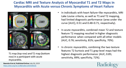
Background The establishment of a timely and correct diagnosis in heart failure-like myocarditis remains one of the most challenging in clinical cardiology.PurposeTo assess the diagnostic potential of texture analysis in heart failure-like myocarditis with comparison to endomyocardial biopsy (EMB) as the reference standard.Materials and MethodsSeventy-one study participants from the Magnetic Resonance Imaging in Myocarditis (MyoRacer) trial (ClinicalTrials.gov registration no. NCT02177630) with clinical suspicion for myocarditis and symptoms of heart failure were prospectively included (from August 2012 to May 2015) in the study. Participants underwent biventricular EMB and cardiac MRI at 1.5 T, including native T1 and T2 mapping and standard Lake Louise criteria. Texture analysis was applied on T1 and T2 maps by using an open-source software. Stepwise dimension reduction was performed for selecting features enabling the diagnosis of myocarditis. Diagnostic performance was assessed from the area under the curve (AUC) from receiver operating characteristic analyses with 10-fold cross validation.ResultsIn participants with acute heart failure-like myocarditis (n = 31; mean age, 47 years +/- 17; 10 women), the texture feature GrayLevelNonUniformity from T2 maps (T2_GLNU) showed diagnostic performance similar to that of mean myocardial T2 time (AUC, 0.69 for both). The combination of mean T2 time and T2_GLNU had the highest AUC (0.76; 95% confidence interval [CI]: 0.43, 0.95), with sensitivity of 81% (25 of 31) and specificity of 71% (22 of 31). In patients with chronic heart failure-like myocarditis (n = 40; mean age, 48 years +/- 13; 12 women), the histogram feature T2_kurtosis demonstrated superior diagnostic performance compared to that of all other single parameters (AUC, 0.81; 95% CI: 0.66, 0.96). The combination of the two texture features, T2_kurtosis and the GrayLevelNonUniformity from T1, had the highest diagnostic performance (AUC, 0.85; 95% CI: 0.57, 0.90; sensitivity, 90% [36 of 40]; and specificity, 72% [29 of 40]).ConclusionIn this proof-of-concept study, texture analysis applied on cardiac MRI T1 and T2 mapping delivers quantitative imaging parameters for the diagnosis of acute or chronic heart failure-like myocarditis and might be superior to Lake Louise criteria or averaged myocardial T1 or T2 values.